Bone Cross Section - Compact Bone Cross-Section - Printable. Smartdraw includes 1000s of professional healthcare and anatomy chart templates that you can modify and make your own. Section of bone marrow affected by myeloma seen under a microscope. Internal structure of a human long bone. The outlined area is a cross section of an osteon of compact bone. Vector illustration scheme of bone cross section.
Vector illustration scheme of bone cross section. The upper (biting) surfaces of the tooth are at top, with the lower sections (bottom) embedded in the gums and jaw bone (not shown). There are trabeculae in spongy bone which gives its sponge like appearance. To the left is muscle tissue, and to the right is bone marrow. Compact bone is the denser, stronger of the two types of bone tissue ( (figure) ).
Dinosaur Bone Cross Section | Osteon remains of this bone ar… | Flickr from c1.staticflickr.com Browse 4,294 bone cross section stock photos and images available, or search for human bone cross section to find more great stock photos and pictures. Find the perfect bone cross section stock photos and editorial news pictures from getty images. Information from its description page there is shown below. Neurons, grey matter with motor neuron cell bodies, white matter with myelinated nerve fibers. In the center of each osteon is the central canal, a space that houses blood vessels and nerves that supply bone. Size of this png preview of this svg file: Concentric layers of bone cells (osteocytes) and bone matrix surround the central canal. By printing out this quiz and taking it with pen and paper creates for a good variation to only playing it online.
Smartdraw includes 1000s of professional healthcare and anatomy chart templates that you can modify and make your own.
Commons is a freely licensed. Section of bone marrow affected by myeloma seen under a microscope. The choice of ct versus mr depends on the structures and the disease processes that require assessment, delineation, and characterization. Sections of bone marrow tissue. Smartdraw includes 1000s of professional healthcare and anatomy chart templates that you can modify and make your own. To the left is muscle tissue, and to the right is bone marrow. Internal structure of a human long bone, with a magnified cross section of the interior. Two types of bone tissues in cross section of a long bone : Browse 53 bone marrow cross section stock photos and images available, or search for bone cross section or bone cells to find more great stock photos and pictures. Find the perfect bone cross section stock photos and editorial news pictures from getty images. While it is not as hard as compact bone, spongy bone plays an important role of protecting the marrow where blood cells are produced. An outer 'fibrous layer' containing mainly fibroblasts, and an inner 'cambium layer' containing progenitor cells. Two types of bone tissues in cross section of a long bone :
Foot bone anatomy x ray 12 photos of the foot bone anatomy x ray foot bone anatomy x ray, bone, foot bone anatomy x ray. Diagram with articular cartilage, marrow, spongy bone, medullary cavity, endosteum, diaphysis, and periosteum. To the left is muscle tissue, and to the right is bone marrow. Commons is a freely licensed. Muscles and bones in the arm 12 photos of the muscles and bones in the arm muscles and.
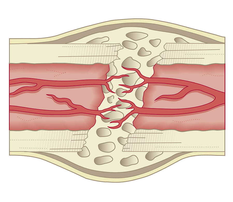
What is a stress fracture? Part 1 of 2 - stay safe! from physioworkshsv.com No need to register, buy now! Concentric layers of bone cells (osteocytes) and bone matrix surround the central canal. Histology sauropod vertebra picture of the week these pictures of this page are about:long bone cross section. An outer 'fibrous layer' containing mainly fibroblasts, and an inner 'cambium layer' containing progenitor cells. The central tubular region of the bone, called the diaphysis, flares outward near the end to form the metaphysis, which contains a largely cancellous, or spongy, interior. Internal structure of a human long bone. Related posts of cross section of human bone diagram foot bone anatomy x ray. A thorough knowledge of the two imaging …
Information from its description page there is shown below.
The choice of ct versus mr depends on the structures and the disease processes that require assessment, delineation, and characterization. Explaned distal and proximal epiphysis. Diagram with articular cartilage, marrow, spongy bone, medullary cavity, endosteum, diaphysis, and periosteum. Concentric layers of bone cells (osteocytes) and bone matrix surround the central canal. For example, if i missed labeling anything, or any parts of the bone are missing. I am not an expert on this subject, so i was wondering if anyone could put their input on this image. This is known as the periosteum. There are trabeculae in spongy bone which gives its sponge like appearance. It can be found under the periosteum and in the diaphyses of long bones, where it provides support and protection. An outer 'fibrous layer' containing mainly fibroblasts, and an inner 'cambium layer' containing progenitor cells. This is a file from the wikimedia commons. Find the perfect cross section bone stock photo. Section of bone marrow affected by myeloma seen under a microscope.
Browse 53 bone marrow cross section stock photos and images available, or search for bone cross section or bone cells to find more great stock photos and pictures. Explaned distal and proximal epiphysis. They are obtained by taking imaginary slices perpendicular to the main axis of organs, vessels, nerves, bones, soft tissue, or even the entire human body. Concentric layers of bone cells (osteocytes) and bone matrix surround the central canal. In the center of each osteon is the central canal, a space that houses blood vessels and nerves that supply bone.
Cross Section Biomedical Illustration Of Bone Repairing Itself With New, Soft, Spongy Callus ... from images.fineartamerica.com Diagram with articular cartilage, marrow, spongy bone, medullary cavity, endosteum, diaphysis, and periosteum. Sections of bone marrow tissue. Smartdraw includes 1000s of professional healthcare and anatomy chart templates that you can modify and make your own. Muscles and bones in the arm 12 photos of the muscles and bones in the arm muscles and. No need to register, buy now! The choice of ct versus mr depends on the structures and the disease processes that require assessment, delineation, and characterization. Section of bone marrow affected by myeloma seen under a microscope. It can be found under the periosteum and in the diaphyses of long bones, where it provides support and protection.
Cross section bone stock photos & cross section bone stock images.
Find the perfect bone cross section stock photos and editorial news pictures from getty images. Select from premium bone cross section of the highest quality. Browse 4,294 bone cross section stock photos and images available, or search for human bone cross section to find more great stock photos and pictures. The outlined area is a cross section of an osteon of compact bone. Beautiful tooth cross section illustration, deep blue background and sparkling lights around. Commons is a freely licensed. Diagram with articular cartilage, marrow, spongy bone, medullary cavity, endosteum, diaphysis, and periosteum. An outer 'fibrous layer' containing mainly fibroblasts, and an inner 'cambium layer' containing progenitor cells. No need to register, buy now! By printing out this quiz and taking it with pen and paper creates for a good variation to only playing it online. I am not an expert on this subject, so i was wondering if anyone could put their input on this image. Information from its description page there is shown below. Smartdraw includes 1000s of professional healthcare and anatomy chart templates that you can modify and make your own.

0 Comments:
Posting Komentar